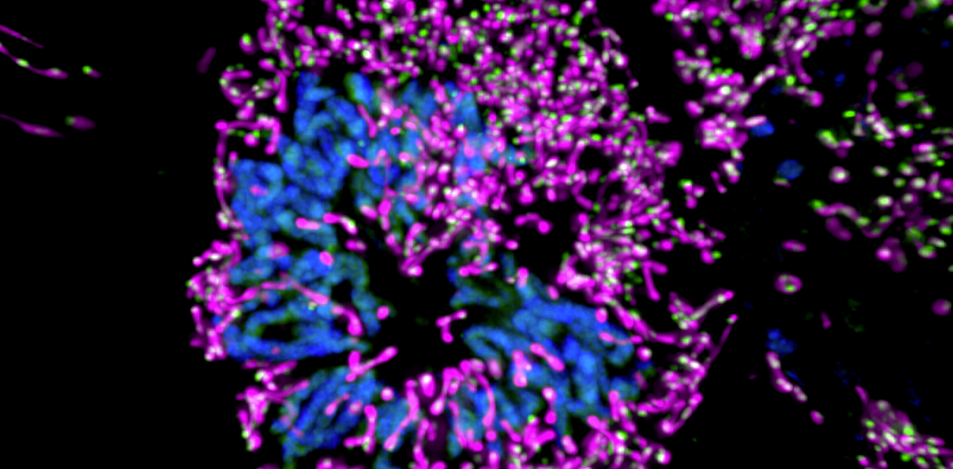
This human cell was obtained by fluorescence microscopy, to distinguish it Nucleus In blue, it’s mitochondria in purple and her nucleoid in green. This image (taken from a searchable video here) shows the complexity of the cell and its components needed to transmit genetic information. This knowledge is necessary to suggest treatments for certain genetic diseases.
Let’s define: the nucleus seals the DNA, the millions of genetic sequences that can be compressed and form chromosomes (in blue). These sequences carry the essential information for the synthesis of our cellular components, the genes.
However, the nucleus is not the only one that hosts genetic information: hundreds of copies of small circular DNA are found in the core of mitochondria (in purple), which are the energy centers of cells. These rather long, tube-shaped membranous structures, which are often interconnected, have their own genetic material, mitochondrial DNA (in green). This mitochondrial genome, which takes the name nucleoid when the proteins associated with it are taken into account, carries quite a few genes. But they are essential to the energy-producing machinery necessary for the functioning of cells (and outside the body). So it is impossible to do without it!
Thus, severe neuromuscular diseases, myopathy and neurodegenerative disorders Mitochondrial DNA sequence mutations or nucleotide disappearances
Normally, when mitochondrial DNA undergoes mutations, the damaged mitochondria are isolated from the others, and then discarded by mitochondrial QoS: this is called mitophagy. Otherwise, mutations accumulate: when the level of mutated mitochondrial DNA exceeds the level of healthy mitochondrial DNA, pathology begins, and clinical symptoms appear. It is therefore essential to understand how the cell detects and activates this exclusion. In Angers, within our team Metolabwe explore the pathology of mitochondrial dynamics to better understand the defects of mitochondrial DNA maintenance.
Mitochondria, dynamic structures
Indeed, fluorescent microscopy data showed that mitochondria are highly dynamic structures, constantly being remodeled by fission, fusion and shape-shifting events. Thus mitochondrial dynamics controls the space between each nucleoid and allows its homogeneous distribution within the mitochondria.
Moreover, the mitochondria themselves are mobile and move around the cell. Thus mitochondrial dynamics ensures the exchange of energy content and puts the energy production mechanism into use. To illustrate, let’s take the example of a neuron, a cell of the nervous system with a very large extension whose end transmits a signal to the next neuron. Mitochondria that are produced in the cell body must migrate in these long stretches to ensure energy is produced at the tip. At the cellular level, mitochondria are not the only structures that move and remodel themselves: transmembrane vesicles, responsible for ensuring quality control of components, also patrol in search of defective elements.
Observing intracellular mitochondrial behavior or nucleoid distribution in different mutant mitochondrial models allows us to perform tests to improve mitochondrial quality control and disease prevention. Thus we are studying cells from patients with mitochondrial disease that present a different percentage of advanced mitochondrial DNA. The efficiency of the system to get rid of damaged mitochondria is evaluated by microscopy techniques with specific markers. Several pharmacological and genetic approaches are being developed to control this quality control system, and to enhance the preservation of mitochondrial DNA.

“Subtly charming problem solver. Extreme tv enthusiast. Web scholar. Evil beer expert. Music nerd. Food junkie.”

